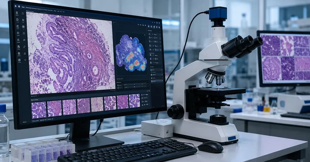
Digital pathology and AI-assisted microscopy are transforming diagnostics by replacing manual microscope workflows with cloud-based, AI-driven tissue analysis. Growing disease burdens, workforce shortages, automation, and regulatory support are accelerating adoption. Advanced AI improves diagnostic accuracy, efficiency, and biomarker discovery, positioning computational pathology as a core pillar of future precision medicine.
The Digital Pathology and AI Assisted Microscopy Market, valued at USD 1.6 billion in 2026, is anticipated to expand at a CAGR of 18.3%, reaching USD 3.7 billion by 2031.
This distinct upward trajectory represents more than a predictable baseline curve of hardware procurement; it marks the late-stage dismantling of an optical monopoly that has dominated medicine since Rudolf Virchow introduced cell theory in 1858. For more than a century, clinical diagnostic validation has depended on the subjective human eye looking through a light microscope lens at a thin slice of chemically treated tissue. However, escalating caseload complexities, shrinking pathologist workforces, and the undeniable mathematical superiority of pixel-level computing are shifting tissue analysis from glass slides to automated cloud infrastructure. This market evolution does not simply digitize manual workflows; it redefines tissue data as a highly structured, scalable digital asset.
The Anatomy of Institutional Adoption
The true catalyst for this projected growth is a profound structural shift across major domestic healthcare networks, academic medical centers, and biopharma consortia. In previous years, digitizing slide volumes was largely constrained by the financial friction of acquiring whole-slide imaging (WSI) hardware, building massive on-premise storage arrays, and navigating complex regulatory gaps. The logistical math has fundamentally flipped. Enterprise health systems no longer view digital pathology as an expensive IT luxury, but as an operational necessity to stave off systemic labor collapse.
A stark reality that dominates the modern laboratory is that the volume of biopsy requests continues to escalate due to an aging population and aggressive cancer screening protocols, while the total number of practicing pathologists is steadily declining. This operational deficit means labs must optimize throughput without introducing diagnostic drift. By shifting the initial triage layer from human observation to deep-learning vision models, labs can systematically separate routine benign cases from highly complex cases that require multidisciplinary tumor board review. This shift dramatically reduces the time spent hunting for minute cellular details across a massive piece of tissue.
Operational Insight: Whole-slide scanners generate exceptionally dense images frequently exceeding 2 to 3 gigabytes per slide at 40× magnification. Storing and managing this volume of multi-resolution, pyramidal digital assets across millions of annual biopsies requires robust enterprise systems. Because of this data density, the underlying software architecture, rather than the mechanical scanner, dictates the long-term economics of modern pathology labs.
Furthermore, structural support from regulatory and reimbursement frameworks has provided necessary market confidence. The introduction of specific Category III CPT codes by the American Medical Association for digital pathology clinical processes, alongside a maturation of FDA clearance pathways for automated diagnostic algorithms, has established a clear monetization roadmap. Hospitals can finally project a tangible return on investment based on clear operational metrics:
Decreased slide retrieval overhead
Faster clinical turnaround times
Significant reductions in expensive external consult shipping fees
For a Tech-Convergence & Product Narrative
Volume of Disease Increasing: The number of people with cancer is growing all around the world. The increased number of people suffering from chronic diseases throughout the world will also contribute to the demand for pathology services, as will the recommendations of health authorities advocating the use of digital pathology and AI microscope applications to increase their ability to diagnose patients faster and more accurately by improving throughput and the speed at which they can identify disease.
Regulatory Agencies Supporting AI in Diagnostics: Government regulatory agencies are establishing guidelines for the future regulatory process for digital pathology and AI technologies. The FDA has implemented the “Digital Pathology Program” to assist with the understanding of validation and use of whole slide imaging and AI algorithms for the diagnosis and monitoring of health conditions. The EU’s Medical Device Regulation (MDR) establishes a framework for the regulation of software and AI as medical devices. This will improve safety for patients and interoperability between devices. Regulatory clarity will lower the barrier to the clinical adoption of digital pathology and AI platforms, which should help promote additional investments.
Patent Activity & New Innovation Incentives: The number of patents filed in the areas of AI-based image analysis, automated microscopy workflow processes, and digital slide annotation is continuing to grow, indicating that innovation is continuing in these areas. The patent activity generated from research institutions, health care consortia, and health technology companies reflects a significant R&D investment and encourages the competition and innovation that will provide laboratories with the next generation of products to improve the speed, accuracy, and ease of interpretive analysis used in clinical practice or research.
Increased Operational Efficiency Via Automation: Digital pathology and AI-enabled microscopy provide automated solutions for many repetitive tasks performed in laboratories, such as cell counting, biomarker quantification, and pattern recognition, producing faster, more accurate results than traditional methods. Automated solutions also provide laboratories with the means to maintain regulatory compliance. Digital pathology and AI tools are integrated into laboratory information systems and electronic health records.
From Vision Automation to Deep Discovery
The technical frontier of AI-assisted microscopy has moved beyond basic geometric pattern matching. Early iterations of image analysis software were brittle, often failing when confronted with routine variances in tissue preparation, slide thickness, or stain intensity from different laboratories. Modern deep-learning models leverage self-supervised learning architectures trained on millions of unannotated whole-slide images, giving them remarkable robustness against routine workflow variations.
These computational engines serve two distinct roles in clinical settings:
1. Workflow Automation and Quality Assurance
At the routine level, algorithms act as an indefatigable second set of eyes. They automate high-volume quantitative tasks that are prone to human fatigue and inter-observer variability, such as counting mitotic figures, tracking tumor-infiltrating lymphocytes (TILs), and calculating immunohistochemistry (IHC) scoring for HER2 or PD-L1 expression. Instead of a clinician manually approximating cell counts across ten distinct high-power fields, an AI assistant evaluates every pixel on the digital slide in seconds, outputting an objective, reproducible metric.
2. Extraction of Novel Sub-visual Biomarkers
The more radical evolution lies in uncovering morphological features that are entirely imperceptible to human vision. Advanced convolutional neural networks (CNNs) can identify spatial relationships within the tumor microenvironment, such as subtle variations in nuclear texture, local cellular density gradients, and architectural ordering that correlate directly with underlying genomic mutations or disease recurrence risk.
Engineers can train models to predict therapeutic responses, microsatellite instability (MSI) status, or specific genetic alterations directly from a standard, inexpensive Hematoxylin and Eosin (H&E) slide. This development could bypass the need to immediately trigger expensive, time-consuming next-generation sequencing assays, allowing clinicians to make rapid, preliminary therapeutic decisions in a fraction of the time.
Strategic Landscape of US Innovators
The domestic market is defined by a dense concentration of specialized technology vendors, enterprise software firms, and traditional medical device manufacturers that are building integrated end-to-end ecosystems. To understand the operational trajectories driving the industry toward its 2031 projection, we must analyze the product developments and strategies of prominent US-based players.
Proscia (Philadelphia, Pennsylvania)
Proscia has established itself as an open, vendor-agnostic enterprise software provider. Recognizing that large medical systems rarely rely on a single hardware manufacturer, the company designed its core platform, Concentriq, as a flexible digital backbone capable of centralizing slides from diverse scanning systems. By decoupling the underlying software orchestration layer from the physical imaging hardware, Proscia circumvents vendor lock-in. Their platform seamlessly integrates third-party and proprietary AI modules directly into the pathologist's daily review environment, streamlining the transition to computational operations.
Paige AI (New York, New York)
Originating from research out of Memorial Sloan Kettering Cancer Center, Paige AI focused heavily on achieving regulatory validation for computational diagnostic tools. The company secured a historic FDA de novo clearance for its primary diagnostic software, Paige Prostate, designed to help identify areas of interest suspect for cancer. Rather than relying on small, highly curated academic datasets, Paige built its foundation models on vast repositories of diverse real-world slides. Their deep-learning models detect subtle focus points of invasive carcinoma that might otherwise be overlooked during manual screening of high-volume biopsy backlogs.
PathAI (Boston, Massachusetts)
PathAI operates at the intersection of high-throughput clinical diagnostics and biopharma drug discovery. Through its PathAI Enterprise platforms and AISight management software, the firm provides robust computational tools to quantify complex tissue phenotypes. A core strategic focus for PathAI is accelerating clinical trials. By standardizing patient stratification metrics and identifying sub-visual tissue responses to novel therapeutics, their technology helps pharmaceutical sponsors reduce ambiguous endpoints in oncology and immunology trials.
Tempus AI (Chicago, Illinois)
Tempus approaches digital pathology through a massive, multi-modal data network. By blending comprehensive clinical tracking, genomic sequencing records, and digitized pathology data, the company treats the whole slide as one piece of a broader diagnostic puzzle. Their Tempus One and specialized digital pathology solutions focus heavily on algorithmic profiling, analyzing standard H&E morphology to identify deep molecular signatures. This integrated framework bridges the historic gap between pure morphology and advanced molecular diagnostics.
The product distributions and operational centers of these major domestic innovators highlight the targeted, solution-driven nature of current commercial development:
Organization | Core Product Platform | Primary Technological Development | Operational Center |
Proscia | Concentriq Enterprise | Vendor-agnostic software orchestration and enterprise image management (Proscia, 2015) | Philadelphia, PA |
Paige AI | Paige Prostate & Paige Breast | FDA-cleared deep learning systems for tissue detection and diagnostic screening | New York, NY |
PathAI | AISight / PathAI Enterprise | Quantitative multi-modal profiling for trial stratification and clinical workflows | Boston, MA |
Tempus AI | Tempus Path | Multi-modal integration of morphology, clinical records, and genomic data | Chicago, IL |
Market Headwinds and Pragmatic Restraints
Despite strong growth projections, treating the widespread adoption of AI-assisted pathology as an inevitability overlooks serious structural hurdles. The path forward is not a simple software rollout; it requires big changes to institutional IT architecture and laboratory economics.
Storage Economics and Infrastructure Strain
The financial strain of data storage remains a persistent barrier for medium and smaller community hospital systems. While cloud computing costs continue to drop, the sheer volume of continuous high-resolution tissue scanning creates long-term storage liabilities.
Laboratories must establish strict data lifecycle management policies:
Deciding when a slide can be safely downgraded to lower resolutions
Determining when it must be archived in deep cold storage
Evaluating how long a slide must remain readily accessible for acute clinical comparison
Interoperability Gaps and Data Silos
True software interoperability is an ongoing battle. While the DICOM (Digital Imaging and Communications in Medicine) standard has made significant progress in paving a uniform data format, many proprietary hardware systems still write data to unique, closed file formats. This friction means IT teams often spend substantial time creating custom pipelines to transport images from a scanner to a third-party AI algorithm, and then back to the central Laboratory Information System (LIS).
Algorithmic Generalization and Training Bias
The most challenging clinical risk centers on algorithmic bias. A tissue model trained exclusively on slides from a premier academic institution using a specific automated stainer may see its accuracy degrade when deployed in a rural clinic using manual staining techniques. The industry must push toward rigorous, multi-site external validation across highly diverse patient cohorts to ensure diagnostic algorithms remain safe and reliable regardless of where the tissue sample was processed.
The Next Decade of Spatial Tissue Intelligence
Looking toward 2031, the market will likely move past individual diagnostic workflows and shift toward full operational integration. We are moving toward a paradigm where a microscope is no longer an isolated analog tool, but an intelligent portal connected to an expansive cloud network.
We can expect a deep convergence of digital pathology with other key diagnostic fields, especially radiology and spatial transcriptomics. Future enterprise imaging systems will likely map a patient's macro-level imaging data, such as a 3D MRI or CT scan, directly to the micro-level cell structures found on a digital biopsy slide. This multi-scale approach will provide oncology care teams with an uncompromised view of disease progression.
At the same time, the rise of "label-free" optical microscopy techniques powered by deep learning could allow labs to bypass traditional chemical tissue processing entirely. By utilizing advanced algorithms to virtually stain fresh, unfixed tissue slices in real time, clinical facilities can radically compress turnaround times from hours to minutes. This evolution will firmly cement computational tissue analysis not just as an optimization tool, but as the foundational architecture for the future of precision medicine.
Key Developments that are driving market growth
In March 2025, Philips Healthcare announced the expansion of its IntelliSite Pathology Solution (PIPS) in collaboration with Ibex Medical Analytics, introducing new AI-powered capabilities designed to enhance diagnostic workflows and address growing pathology workloads. The updated platform supports improved interoperability between digital pathology scanners and advanced AI applications for cancer diagnostics, including case prioritization and automated image analysis. PIPS 6.0 also delivers streamlined workflows for pathology teams, making digital and AI-assisted diagnosis more efficient and scalable in clinical laboratories. This development reflects the company’s broader strategy to accelerate the adoption of AI tools in routine pathology practice worldwide and improve diagnostic confidence and turnaround times.
Have questions?
More Blogs
Top U.S. Healthcare Industry Trends to Watch in 2026 for the Global Healthcare Market
The global healthcare industry in 2026 is shifting toward AI-driven efficiency, stronger cybersecurity, outpatient care expansion, and domestic pharmaceutical manufacturing. U.S. healthcare leaders are prioritizing workforce support, operational resilience, value-based care, and integrated digital systems to improve patient outcomes while controlling costs.
How AI Is Reshaping the American Healthcare Industry
The U.S. healthcare industry is rapidly integrating artificial intelligence across clinical, administrative, and pharmaceutical operations. AI is improving diagnostics, automating workflows, reducing clinician burden, accelerating drug discovery, and enhancing personalized care. With strong adoption by hospitals and tech companies, AI is transforming healthcare delivery while driving the U.S. AI healthcare market toward significant growth by 2031.
Top Companies Leading the Silicon-Based Capacitor Revolution
Introduction: The Rise of Silicon-Based Capacitors: Electronic circuits use basic capacitors as storage devices to handle three essential tasks including filtering, decoupling, and signal coupling. High-performance silicon capacitors h...